Pest & Crop
Newsletter
Purdue Cooperative Extension Service
Purdue Cooperative Extension Service
Issue 22, August 27, 2015 • USDA-NIFA Extension IPM Grant
CLICK HERE FOR A PDF VERSION OF THIS ISSUE ![]()
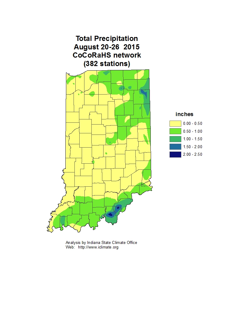
Recently there has been increased interest in utilizing cover crops in our corn and soybean production systems because of government sponsored cost share programs and soil health benefits. Concurrently, there has also been increased utilization of soil residual herbicides in our corn and soybean production systems to help manage herbicide resistant weeds such as marestail, pigweeds, and ragweeds. Soil residual herbicides can remain active in the soil for anywhere from weeks to months after application. The length of time a residual herbicide remains biologically active in the soil is influenced by soil type, soil pH, organic matter, rainfall, and temperature. Since these factors will vary from field to field, definitive time intervals of residual herbicide activity can be difficult to predict.
A significant challenge has arisen because use of residual herbicides in our corn and soybean production systems may interfere with establishment of fall seeded cover crops. An unfortunate coincidence is that many of the crops being used for cover crops were not evaluated for herbicide carryover when field research was being conducted for support of the EPA label of the respective herbicide. As a result, data are lacking regarding rotational intervals for establishment of many cover crop species.
Over the last couple of growing seasons we have established experiments designed to evaluate the impact of commonly used residual herbicides on the establishment of many cover crop species. In addition, our colleagues in adjacent states have conducted similar research and we feel like we have a better handle on this topic now than we did two or three years ago. As was mentioned above, predicting herbicide persistence is complicated because so many different factors can influence herbicide dissipation in the soil.
As a general rule, residual herbicides that have activity on grass weeds can interfere with the establishment of some grass cover crop species, especially the smaller seeded ryegrass species. Residual herbicides from the group 2 (ALS), group 5 (triazine), group 14 (PPO), or group 27 (bleacher) can interfere with the establishment of some of the broad leaf cover crop species.
More specifically we have learned the following:
This summarizes our current knowledge on establishment of cover crops following the use of residual herbicides. The final two things to mention is that if you have questions about specific situations, one way to address the residual herbicide left in a field is to do a bioassay. Simply collect soil from the area you would like to seed the cover crop into and an area with a similar soil type, but no herbicide residue, and plant seed from the cover crop you would like to use. Observe growth for 3 weeks and if the plants look the same in the untreated and treated soil, you should be safe to plant to desired crop. Another consideration if you do not have time to do a bioassay is to plant a cover crop mixture. Cover crop establishment may be more reliable when mixtures of grass and broadleaf species are purchased and planted. Residual herbicides may interfere with establishment of some species in the mix, but have no effect on other species. The use of mixtures may allow one more protection from complete failure due to excessive residues in the soil. It would be important however to be sure that at least one or two of the species in the mixture is tolerant to the herbicides used in a specific field.
Weather conditions were favorable for several different ear rots of corn in Indiana this year. Currently, Diplodia ear rot has been found most frequently in the state, and as we prepare for harvest, it is important to identify fields that may have ear rots to ensure timely harvest and proper storage of moldy grain.
Diplodia ear rot is caused by the fungus Stenocarpella maydis, and is very common in cornfields across the Corn Belt. This fungus survives in residue and infects plants during and after pollination. Humid weather and rains prior to and after pollination will favor disease development. Diplodia ear rot is identified by white fungal growth on the cob, often forming a mat of fungus across the ear (Figure 1). Infected kernels may also be brown-gray in appearance (Figure 2). Small, black fungal structures called pycnidia may form on the kernels or the cob (Figure 3). The fungus is reported to produce several mycotoxins in South America and South Africa, however, no reports of toxic effects of grain on livestock or humans due to Diplodia ear rot have been reported in the United States. Grain dockage may still occur, however, due to moldy grain.
Purdue University is conducting research on the fungus that causes Diplodia ear rot in hopes of improving management options available to farmers. If you have fields with suspected or confirmed Diplodia ear rot, please email Paty Romero at mromerol@purdue.edu.
Diplodia ear rot may be confused with other ear rots, such as Gibberella ear rot. A different fungus causes each of these rots, and the environmental conditions at and just after silking influence which ear rot may be problematic in a given year.
Gibberella ear rot, caused by the fungus Fusarium graminearum, is common during cool, rainy years. The fungus infects during early silking and pollination, and is favored by cooler temperatures than Diplodia ear rot. This fungus produces a fungal mat on the ear, similar to Diplodia ear rot, but often with a pink or reddish color to the mold (Figure 4). Gibberella zeae produces the mycotoxin deoxynivalenol (DON), commonly referred to as vomitoxin. This mycotoxin can be extremely harmful to swine, and is carefully regulated according to the FDA.
Preventative management of ear rots is critical, and can be accomplished by selecting less susceptible hybrids and reducing the amount of corn residue that can serve as a source for the fungus to overwinter through crop rotation and tillage. In-season management of ear rots is limited at this point, with few fungicides and anti-fungal products available for specific ear rots. Purdue research indicates that currently available fungicides do not have efficacy against these diseases.
Farmers should scout fields prior to harvest and determine the level of incidence of any ear rot in the field. If ear rots are observed in a field, affected areas should be harvested early and grain segregated to avoid mixing moldy and/or mycotoxin contaminated grain with high quality grain. Silage and grain harvested with suspected ear rots should be dried to below 15% moisture. If grain or silage (with kernels present) is kept above this moisture content, mycotoxin can continue to accumulate in grain. All grain contaminated by any ear rot fungus should be stored separately from good grain, and if stored long term, stored below 13% moisture to prevent further growth of fungi. All moldy grain should be tested to determine the presence and level of mycotoxins prior to use in livestock feed.
For more information on these ear rots and managing grain, please see www.cornmycotoxins.com
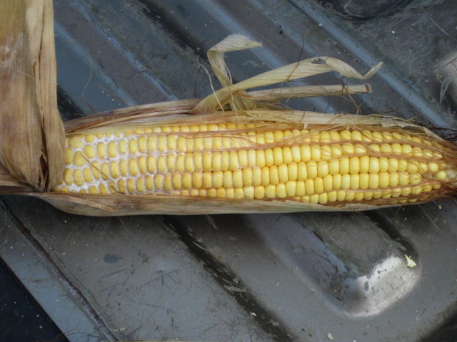
Figure 1. The fungus that causes Diplodia ear rot produces a white fungal mat on the cob.
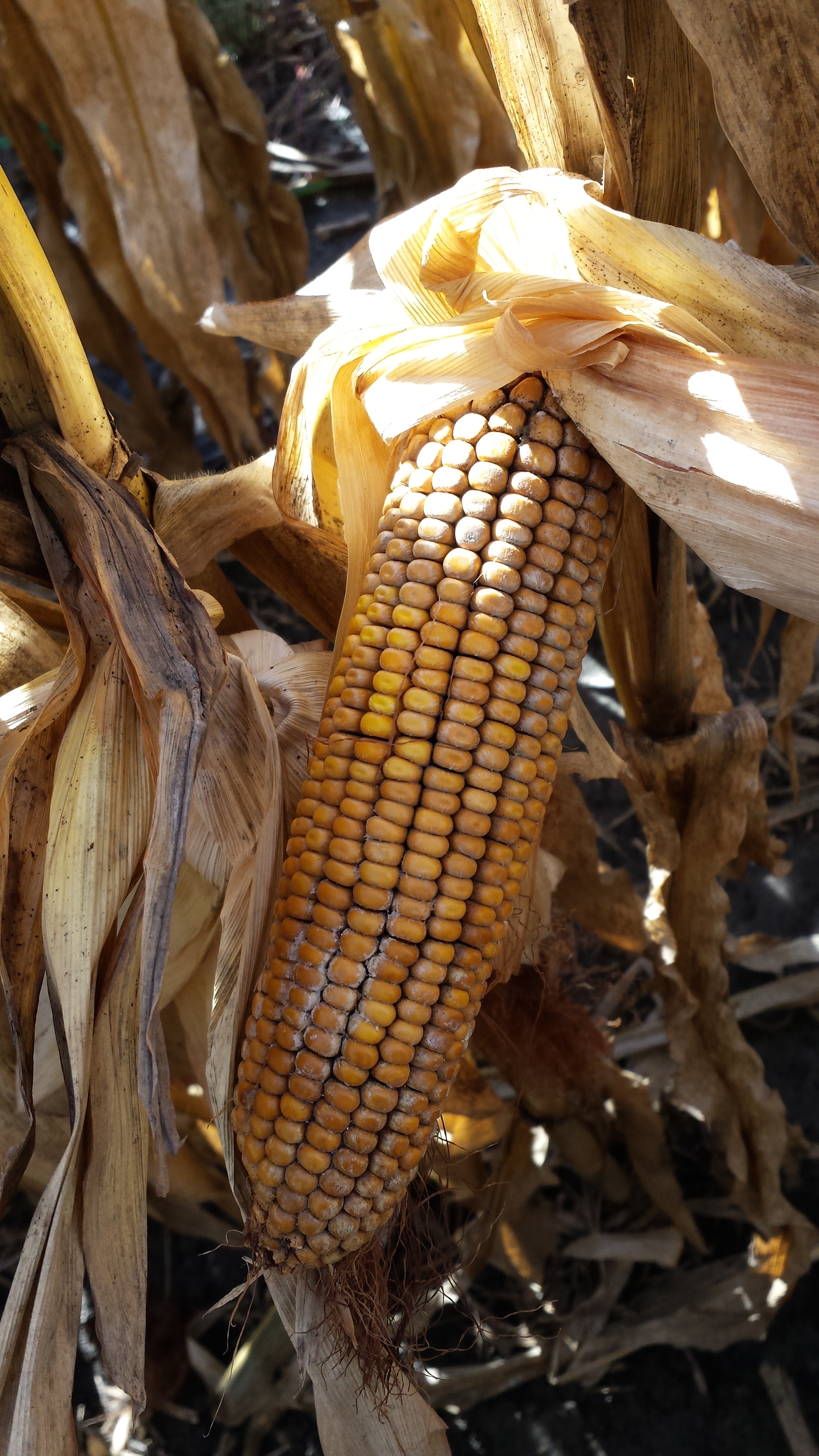
Figure 2. Brown discoloration on ears can also indicate presence of Diplodia ear rot.
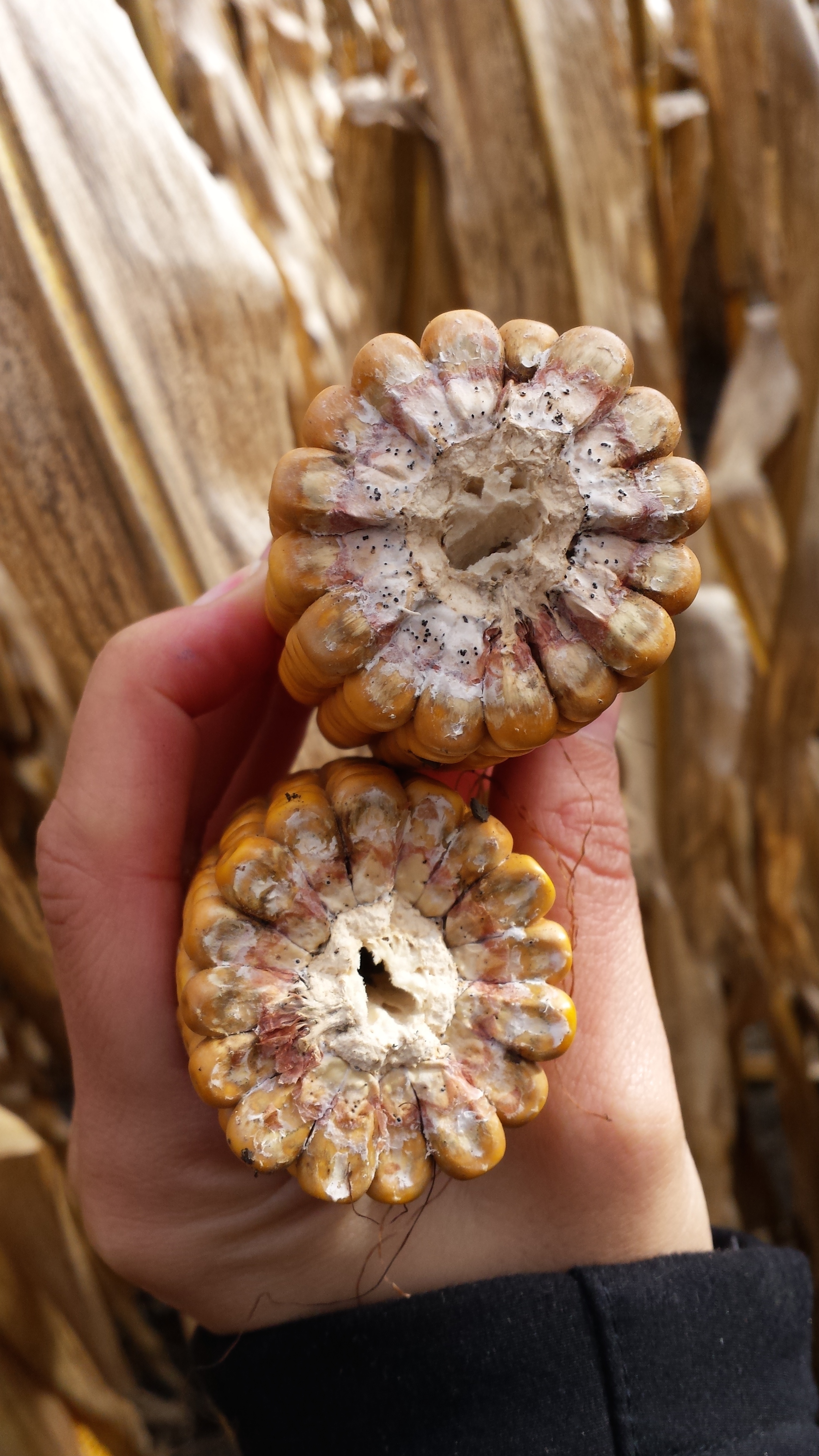
Figure 3. Black fungal structures, called pycnidia, are visible in cob tissue as a result of infection by the fungus that causes Diplodia ear rot.
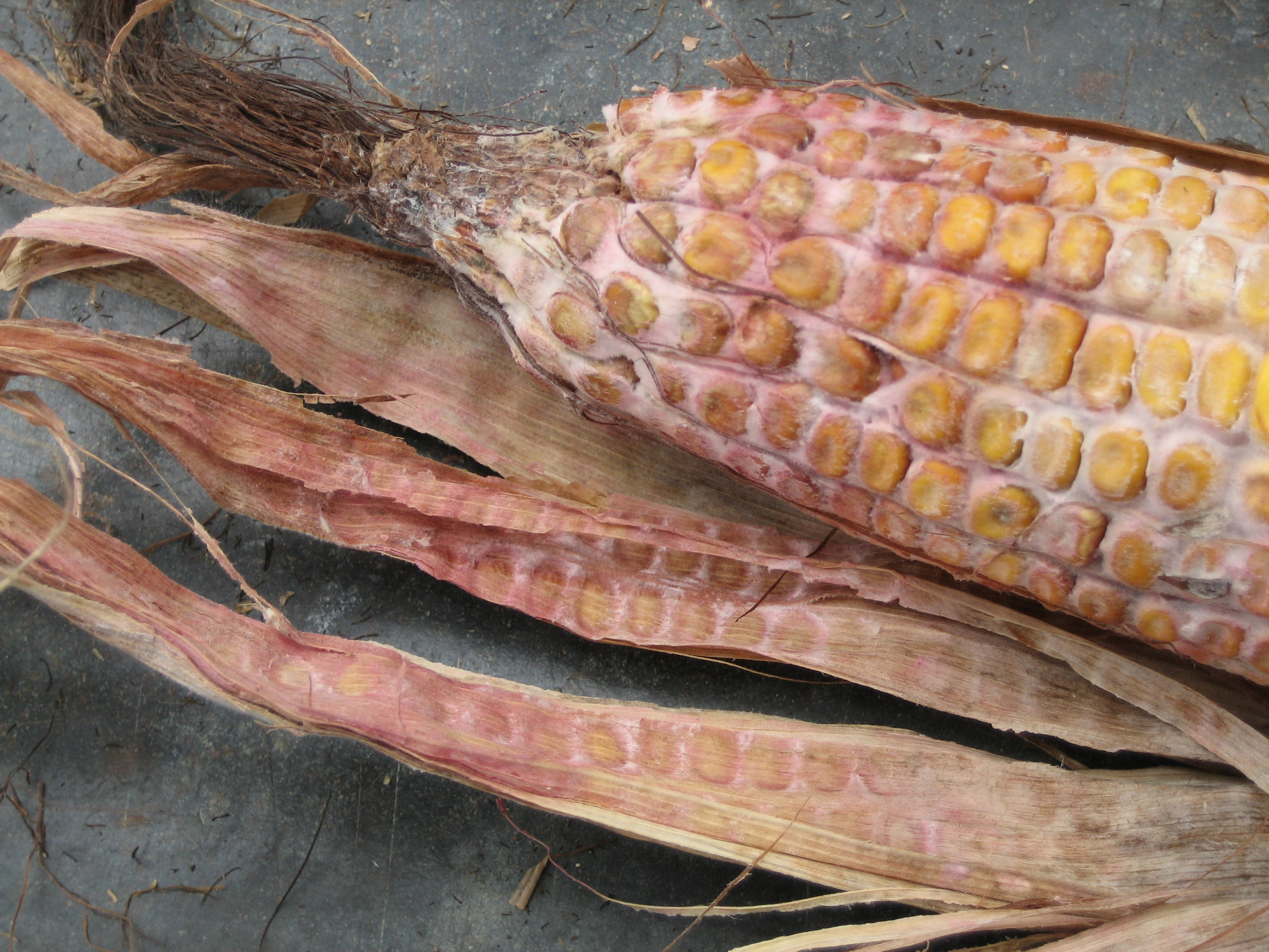
Figure 4. A pink mold on the ear is a sign of the fungus that causes Gibberella ear rot (picture courtesy Charles Woloshuk).
Goss’s wilt of corn, caused by the bacterium Clavibacter michiganensis subsp. nebraskensis, has been confirmed in several fields in northeast Indiana in 2015, making these the most eastern confirmations of the disease in the state to date (Figure 1). The disease has also been detected in northwest Indiana this year.
The bacterium that causes the disease survives in residue and some grassy weed species and infects corn plants through wounds. The diagnostic symptoms of Goss’s wilt include water-soaked lesions that have black “freckles” or specks on or surrounding the lesions (Figure 2). These lesions can be on any leaves of the plant and may be quite large on susceptible hybrids, often causing a scorched leaf symptom from the tip to the base of leaves. (Figure 4) Bacteria can also move onto the leaf surface and give the leaves a shiny appearance (Figure 3).
These lesions are very easy to confuse with the leaf burning and firing that is common across Indiana due to nitrogen deficiency and plant stress. Goss’s wilt foliar lesions may also be easily confused with the fungal disease northern corn leaf blight.
It is important to remember that this disease MUST be diagnosed with at least two methods: microscopic observation of bacterial streaming from symptomatic tissue, and confirmation of the causal bacteria using organism-specific tests. In-field testing is possible with an immunostrip test marketed by the company AgDia (http://www.agdia.com/). However, there are limitations to these strips, and it is not recommended to rely solely on a diagnosis based on these strips. Sample contamination can occur in the field, and strips may occasionally detect the bacterium that causes Goss’s wilt when it is not actually present in the plant. Because of the potential for these false positives, it is important that any sample that tests positive in the field for Goss’s wilt be sent to a diagnostic lab for additional microscopic observation. (www.ppdl.purdue.edu) Plants that are not infected with the bacterium will not exhibit this bacterial streaming, and Goss’s wilt can be ruled out.
It is important to scout fields in these areas to determine if fields are affected by Goss’s wilt because this disease must be managed preventatively. Fields that are affected by Goss’s wilt should be planted to a resistant hybrid in future years.
Other management options include tillage and rotation to help reduce the bacteria population present in the field for the subsequent corn crop. Residue can harbor bacteria for years. In-season management is limited and may not be warranted depending on the growth stage and yield potential of the crop. Fungicides are not recommended to use for management of Goss’s wilt since it is a bacterial disease.
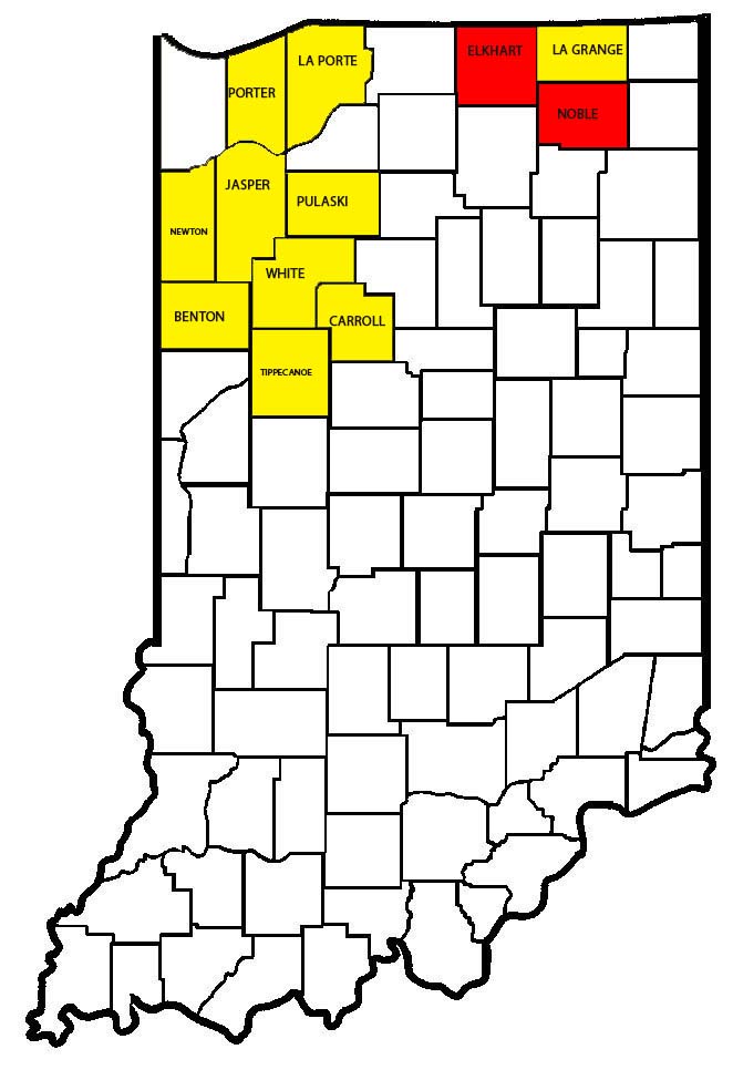
Figure 1: Map showing distribution of Goss’s wilt in Indiana. Counties highlighted in red indicate new confirmations of Goss’s wilt in areas that the disease had not previously been reported. Yellow counties indicate where the disease has been confirmed since 2008.
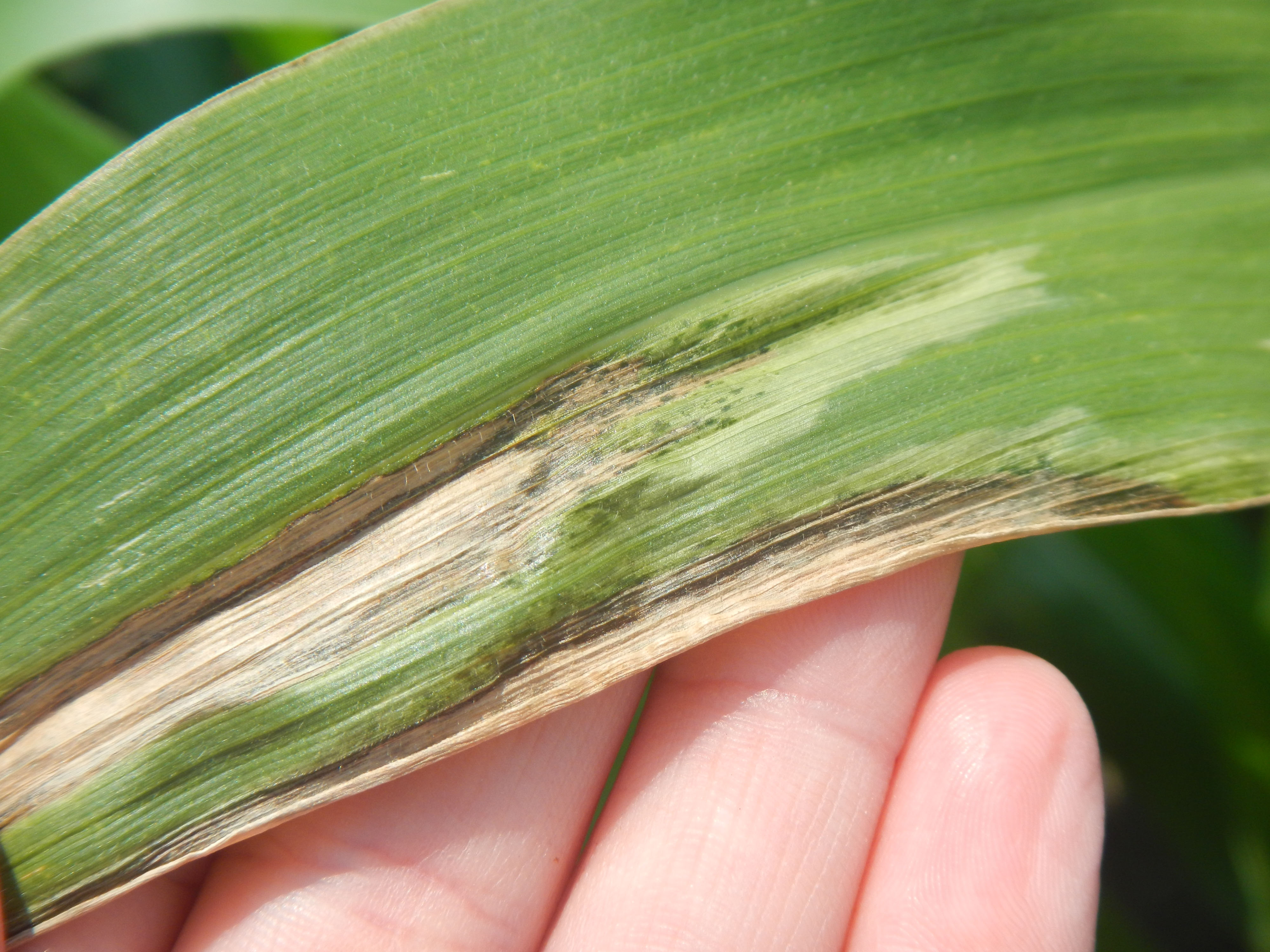
Figure 2: Diagnostic symptoms of Goss’s wilt include water-soaked lesions with black “freckles” or specks on or surrounding the lesions
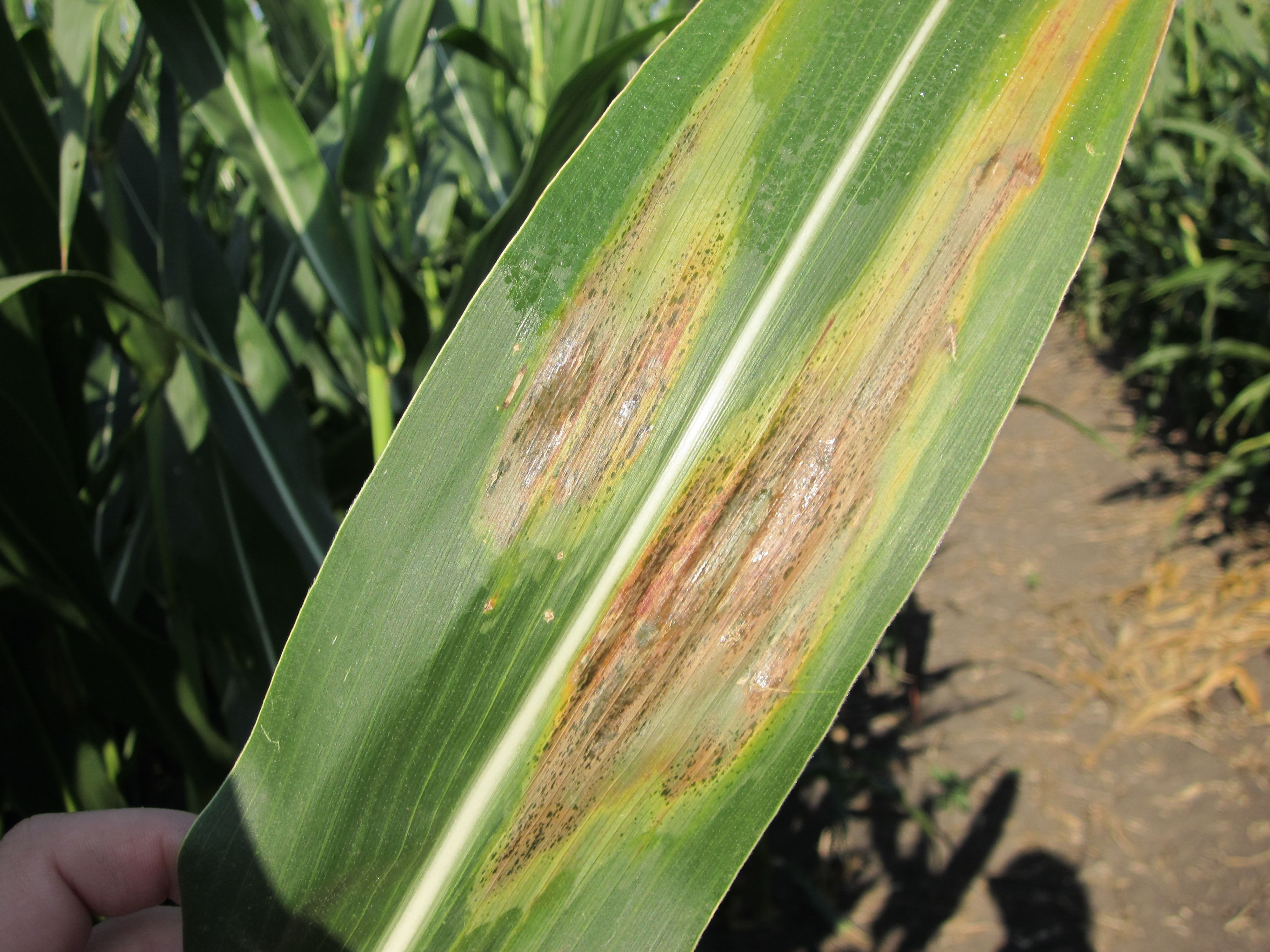
Figure 3: Bacteria on the leaf surface give the leaves a shiny appearance
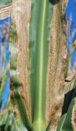
Figure 4: Leaf scorch caused by coalescing Goss’ Wilt lesions
Purdue Cooperative Extension Service
Purdue Extension Entomology
901 W. State Street
West Lafayette, IN, 47907
(765) 494-8761
luck@purdue.edu
@PurdueExtEnt
PurdueEntomology
![]()
If you would like to be alerted by e-mail when the current issue of the Pest&Crop is available on-line, please enter your e-mail address and click the submit button.
It is the policy of the Purdue University Cooperative Extension Service that all persons have equal opportunity and access to its educational programs, services, activities, and facilities without regard to race, religion, color, sex, age, national origin or ancestry, marital status, parental status, sexual orientation, disability or status as a veteran. Purdue University is an Affirmative Action institution. This material may be available in alternative formats.